
Artificial intelligence has quietly moved into the dental chair, and patients around Westford are starting to hear the term at routine visits. AI dental diagnostics means dental imaging software reviews your X-rays and scans and flags anything that looks abnormal, giving your dentist a second set of eyes before treatment decisions get made. The software does not diagnose you. It supports the dentist who does.
What AI Dental Diagnostics Means in Plain English
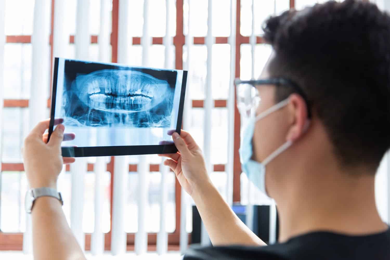
Every dental exam produces images: bitewings, periapical X-rays, sometimes a panoramic image or a 3D scan. For decades, a dentist read those images by eye, using training and experience to separate normal anatomy from early trouble. That is still true today, but the reading is now often paired with software trained on very large collections of dental images.
That software uses machine learning, which means it improves at pattern recognition by processing many examples rather than following a fixed set of instructions written by a programmer. When your image is uploaded, the software compares what it sees against those patterns and highlights areas that deserve a closer look. Your dentist then reviews both the image and the software’s flags, applies clinical judgment, and decides what happens next.
The practical takeaway for patients is simple: nothing about your appointment changes very much. You still sit in the chair, you still get imaged, and you still get a conversation about what your dentist found. What changes is how much support sits behind that conversation.
How the Software Reads Your Dental Images
Different platforms work with different image types, and a single practice may use more than one. The categories below cover what current dental AI tools analyze:
- Intraoral X-rays, including the bitewing and periapical images taken during a standard checkup
- Panoramic X-rays, sometimes called OPGs, which capture a wider view in one exposure
- CBCT scans, the three-dimensional imaging used when a case needs more depth than a flat image can provide
Some platforms process images in real time as they are captured, scanning for caries, bone loss, and other abnormalities as the image loads. Others run analysis after the fact and present findings alongside the original image. In both cases, the output is a set of highlighted areas and supporting detail, not a finished diagnosis.
2D and 3D Images: What the Software Handles
Two-dimensional and three-dimensional images are not interchangeable, and the depth of AI analysis can differ between them. Dental AI systems may analyze intraoral X-rays, panoramic X-rays, and CBCT images to help identify areas that may need closer attention. Some systems can review a range of conditions across both 2D and 3D images, depending on how the software is designed.
These capabilities describe what an AI system is built to assess, not a guarantee about what it will find in every patient’s images. The dentist still reviews the images and uses clinical judgment to determine what the findings mean for your care.
| Image type | What the AI software reviews |
|---|---|
| Intraoral X-rays | 2D analysis, with vendor-reported detection across more than 40 conditions |
| Panoramic X-rays (OPGs) | 2D analysis of a wider single-exposure image |
| CBCT (3D) | 3D analysis, with vendor-reported detection across more than 60 conditions |
What AI Can Help Flag Earlier
The case for this technology rests on timing. Research published in 2024 found that AI algorithms can help dental professionals detect caries, periodontal disease, and oral cancer at an earlier stage. A separate 2025 review reported that AI lets dental radiologists read and interpret radiographs more accurately, improving diagnostic accuracy, and that surgical accuracy and outcomes improve as well.
Patient-facing summaries of the same research describe AI identifying early-stage decay, subtle bone loss patterns, and changes that are easy to overlook on a quick visual pass. Those are exactly the findings that matter most, because small problems caught early tend to need smaller solutions.
It helps to understand why software can catch things an eye might miss. A dentist scanning an image is looking at dozens of teeth and supporting structures at once, under time pressure, in a room with a patient waiting for answers. Software does not get tired, does not get distracted, and applies the same consistent check to every image. That consistency is the value, not magic.
Does AI Replace Your Dentist?
No, and reputable platforms are clear about this. Software can highlight a suspicious area, but it cannot palpate a tooth, check how a restoration is seating, ask you whether that molar has been sensitive to cold, or weigh your medical history against a treatment plan. It also cannot tell you what to do about what it finds.
Think of the relationship as layered review. The software performs a consistent first pass. Your dentist performs the clinical exam, interprets the findings in context, and makes the call. If the two disagree, the dentist’s judgment wins, because the dentist is the one treating a person rather than an image. AI is a tool inside a diagnostic process, not a replacement for the professional running it.
What the Numbers Say
Dental AI has moved past the experimental stage. FDA-cleared dental AI systems are now being used in clinical settings, with research and real-world adoption continuing to grow. Because performance and adoption figures can vary between products and sources, vendor-reported numbers should be viewed as product-specific claims rather than independent benchmarks.
Academic interest has grown alongside commercial adoption. A 2025 paper on transforming dental diagnostics with artificial intelligence has been cited dozens of times, and a 2024 review on optimizing oral and dental diagnoses with AI has drawn similar attention. Teaching tools exist as well, including free AI-powered learning resources for dental students and universities built around real X-ray data. When a technology appears in both clinical settings and education, it shows that AI is becoming an increasingly established part of modern dental diagnostics.
How This Fits Into a Visit at a Westford Dental Office
Cornerstone Dental Group is a private boutique practice in Westford, Massachusetts, and AI diagnostics are part of how the office approaches exams. Digital scanning with iTero technology sits alongside that imaging work, so records are captured digitally rather than through goopy impression trays. Patients from Lowell, Chelmsford, Nashua, Tyngsborough, Acton, and Ayer are all within reasonable reach of the practice.
What does that mean for a checkup? Your images get captured, the software reviews them, and your dentist walks you through what stood out. If the software flags something and the clinical exam confirms it, you get a clear explanation of what it is and what your options are. If the software flags something that turns out to be harmless anatomy, your dentist tells you that too. Good diagnostics include ruling things out, not just finding things.
Questions Worth Asking at Your Next Visit
If you are curious about how your dental office uses this technology, ask. Most dentists are happy to explain their equipment. A few useful questions:
- Which imaging software is used to review my X-rays?
- Can you show me what the software flagged and point it out on the image?
- How does what the software found change my treatment options, if at all?
- How is image analysis handled in terms of billing for my visit?
On that last point, cost and coverage vary by practice and by plan, and there is no single published answer that applies everywhere. The front desk at your dental office is the right place to get specifics for your situation.
The Bottom Line for Patients
AI dental diagnostics is best understood as a consistency tool. It gives your dentist a systematic review of every image, every time, and it can surface early decay and subtle bone changes that are easy to miss on a fast visual pass. It does not diagnose you, it does not replace the exam, and it does not decide your treatment. It gives the person making those decisions better information to work with, which is a reasonable thing to want from any piece of technology in a dental office.
Frequently Asked Questions
Can AI detect problems my dentist might miss?
Research summaries suggest it can help. Studies indicate AI can identify early-stage decay, subtle bone loss patterns, and other findings that are easy to overlook during a standard visual review. The software applies the same systematic check to every image, which reduces the chance that something small gets missed on a busy day. Your dentist still confirms anything the software flags.
Is AI dental diagnostics the same as getting an X-ray?
No, they are two separate steps. The X-ray or scan captures the image, and AI software analyzes that image afterward or in real time as it loads. You do not need different preparation for the AI portion. If your dentist has already decided you need imaging, the analysis is simply part of how those images get reviewed.
Is AI dental diagnostics new?
It has become far more common in recent years. Research papers on the topic appeared in 2024 and 2025, and FDA clearance exists for dental AI products used in clinical settings. What felt experimental a few years ago is becoming a more established part of modern dental diagnostics.
Will my dental insurance cover AI-assisted image analysis?
Coverage and billing for image analysis vary by plan and by dental office, and there is no universal rule that applies to everyone. The most reliable answer comes from your own dental office and your insurance carrier. Ask how image review is handled for your visit before treatment begins so there are no surprises afterward.
The technology will keep improving, and the vocabulary around it will keep shifting. What will not change is the goal: catching problems while they are small, explaining them clearly, and letting you decide what to do next with a dentist who knows your history. That combination of software consistency and human judgment is what makes modern dental diagnostics work.