
A small change on a dental X-ray is easy to overlook. A faint shadow between two teeth, a slight difference in bone height, a hint of something forming just under the surface of a molar. These are the findings that reward a second look, and a second look is exactly what AI-assisted imaging is built to provide.
At practices such as Cornerstone Dental Group in Westford, MA, AI dental diagnostics add a software layer to the radiographs and images a patient already has taken. The software reviews those images, highlights areas that may deserve closer attention, and hands the results back to the dentist. The appointment does not become impersonal or automated. The goal is simpler than that: make sure nothing small slips past unnoticed.
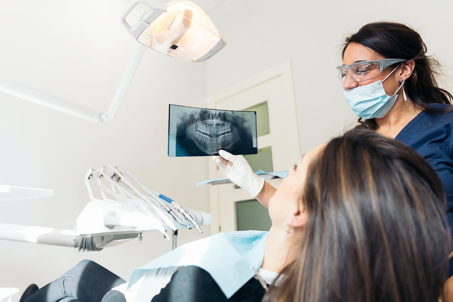
What AI Dental Diagnostics Actually Do
AI-driven diagnostic tools are changing the way dental professionals detect, analyze, and treat oral health issues. In day-to-day practice, that means software that reads the radiographs your dental team already captures and applies deep learning to look for patterns a human eye might skim over. The system marks areas of interest. The dentist then reviews those marks alongside everything else they know about your mouth, your history, and your symptoms.
Published overviews of this technology describe AI systems analyzing X-rays and images to help dentists diagnose oral health conditions more accurately. The wording matters here. The software assists. It does not diagnose on its own, and it does not decide what happens next. A licensed dentist still reads the film, confirms or dismisses what the software flagged, and makes the call.
That division of labor is what makes the technology useful rather than threatening. You get a tool that never gets tired or distracted, paired with a clinician who understands context, comfort, and long-term goals.
Where the Software Sits in the Workflow
Nothing about an AI-assisted exam requires a dramatic change to your visit. The usual sequence still applies, with one extra step tucked into the middle.
- Images are captured the same way they always have been, using the practice’s standard radiograph and scanning equipment.
- The software reviews those images and produces annotations, flagging regions that match patterns it has learned from large sets of prior cases.
- The dentist examines both the original image and the annotated version, comparing what the AI noticed against their own clinical reading.
- The findings get explained to you, often on a screen you can see, so the conversation is about a visual you can follow rather than a description you have to imagine.
Practices that pair AI review with intraoral scanning tools, like the iTero Lumina scanner used for digital impressions, tend to build that same visual language across the whole appointment. The X-ray review, the scan, and the treatment conversation all point at the same screen.
Why Early Detection Is the Whole Point
The value of catching something early is not abstract. It shapes how much treatment you need, how long it takes, and how much of your year gets organized around dental appointments.
Smaller Findings Usually Mean Simpler Treatment
A problem identified while it is still small is generally easier to manage than one discovered after it has grown, spread, or started causing pain. This is true in dentistry as much as anywhere else in healthcare. The earlier the conversation starts, the more options stay on the table, including the option to watch an area and monitor it rather than intervene immediately.
AI review supports that early conversation because it reduces the chance of a subtle change going unmentioned from one visit to the next. A finding that shows up clearly in an annotated image is a finding that gets discussed, documented, and tracked.
Better Images Make Better Conversations
Patients understand their own diagnoses far better when they can see what the dentist sees. Case reports on AI in dentistry describe how AI-enhanced radiographs and visual tools help patients grasp their diagnosis, which in turn supports more informed decisions about treatment. If a picture is genuinely clear, questions get asked sooner and answered more directly.
That matters for anxious patients in particular. Uncertainty tends to feed worry. Seeing the actual image, with the relevant area pointed out, replaces guesswork with something concrete.
The Technology Behind Smart Dental Imaging
The systems now appearing in dental offices did not appear overnight. They sit on years of research into machine learning applied specifically to dental imaging.
Deep Learning and Radiograph Annotation
AI-powered dental imaging uses trained models to review dental X-rays and identify patterns or areas that may deserve a closer look. The software can annotate images, highlight areas of interest, and return those findings quickly enough to support the dentist during the visit. The dentist then reviews the original images alongside the AI-generated annotations as part of the overall examination.
This is not limited to experimental research anymore. AI-assisted imaging is becoming more common in dental care as practices adopt technology that can support image review and help identify subtle findings. For patients, the practical benefit is an additional layer of analysis that can make dental imaging easier to review and discuss.
Building the Data Foundation
Academic research is helping move AI in dentistry forward by creating and organizing large datasets that can be used to develop and improve diagnostic applications. These datasets give AI systems more examples to learn from, helping researchers explore better approaches to image analysis, treatment planning, and other areas of dental care.
Large, well-structured datasets are an important part of developing reliable diagnostic models. As the technology continues to mature, better training data can help AI systems identify patterns more consistently while still keeping the dentist responsible for reviewing the findings and making clinical decisions.
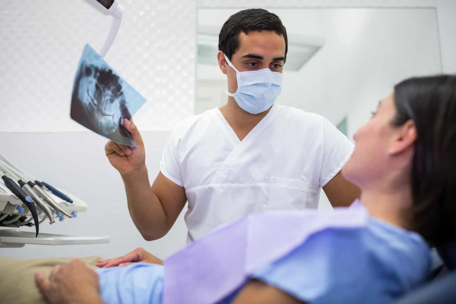
What an AI-Assisted Visit Looks Like in Westford
For a patient in Westford or a neighboring town like Lowell, Chelmsford, Nashua, Tyngsborough, Acton, or Ayer, an AI-assisted exam usually feels like a well-organized regular exam. You sit down, the images get taken, and then the dentist walks through what the software highlighted and what it means for you.
The conversation tends to be practical. Which areas are stable and can simply be monitored. Which areas need a closer look. What a finding might mean if it stays the same, and what it might mean if it changes. Because the annotations are visible on screen, you are not being asked to take anyone’s word for it.
Patients who want a practice with this kind of technology in the room can simply ask. Most dental offices are happy to explain what imaging software they use, who reviews the output, and how results get shared with patients.
Beyond Detection: Planning, Implants, and Smile Design
Diagnostics is only one application. The same class of tools now stretches from smarter diagnosis and radiograph analysis into treatment planning, implant planning, and smile design. That range matters if you are considering anything beyond routine care.
Planning a restoration or an implant involves questions about bone, spacing, and bite that are difficult to answer from a flat film alone. Digital workflows let the dentist model the situation, test an approach before committing to it, and show you the reasoning behind a recommendation. In cosmetic cases, the same digital tools help align expectations with what is realistically achievable.
AI does not make those decisions. It gives the dentist more information to make them with, and gives you more to react to when you are deciding what you want.
What AI Dental Diagnostics Cannot Do
It helps to be clear about the limits, because overstated claims about AI do patients no favors.
- It cannot replace a clinical exam. Looking, probing, and asking questions still surface things an image never will.
- It cannot account for your history on its own. Prior treatment, medications, symptoms, and lifestyle all shape what a finding means for you specifically.
- It cannot make the treatment decision. The dentist interprets the output and recommends a path.
- It cannot substitute for the conversation. Understanding your options is a discussion, not a printout.
The right way to think about AI in dental imaging is as a careful second reviewer that never rushes. Valuable, but only one input among several.
Frequently Asked Questions
Does AI replace the dentist’s diagnosis?
No. AI-assisted software analyzes radiographs and images, then flags areas that may warrant a closer look. A licensed dentist reviews those flags against the clinical exam, your history, and your symptoms before reaching any conclusion. Published descriptions of the technology consistently frame it as a support tool for professionals, not an independent diagnostician.
Is AI imaging different from the X-rays I already get?
The images themselves come from the same radiograph and scanning equipment a practice already uses. The AI layer is software that reviews those images and annotates them. You are not undergoing a separate procedure or a different type of scan, and the appointment timeline stays essentially the same.
How do I know whether my dental office uses AI diagnostics?
Ask directly. A reasonable question is whether the practice uses AI-assisted radiograph review, who interprets the output, and how findings get explained to patients. Practices that have adopted the technology are generally glad to describe it, and a clear answer tells you something useful about how the office communicates in general.
Where is dental AI research heading?
Academic research is helping move dental AI forward by creating large, structured datasets for the development of new AI applications. These datasets give researchers more information to work with when improving diagnostic tools, treatment planning, and other areas of dental care. As the technology continues to develop, better training data may help diagnostic models identify patterns more consistently, supporting both preventive care and more complex treatment planning.
Can seeing the image myself actually help?
It often does. Case reports on AI in dentistry note that AI-enhanced radiographs and visual tools help patients better understand their diagnosis, which supports more informed treatment decisions. When the relevant area is pointed out on screen, you can ask specific questions instead of working from a verbal description alone.
If you are due for a checkup in Westford or a nearby community, it is reasonable to expect your dental team to explain what they see and why it matters. AI-assisted imaging makes that explanation easier to give, and easier to follow, which is ultimately the point.